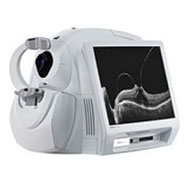
Ocular Coherence Tomography:
A quick non-invasive OCT exam reveals details which help doctors detect potentially debilitating eye diseases, like macular degeneration, glaucoma or diabetic retinopathy, and supports them in making treatment decisions. Doctors have found that early diagnosis and treatment may help to preserve vision.
OCT is a breakthrough in eye care that has allowed doctors to more easily detect eye disorders. The OCT is used to capture detailed images of the eye. The information from OCT images helps your eye doctor to diagnose and manage potentially damaging ocular diseases which, if left untreated, can lead to blindness or seriously impact a person’s quality of life.
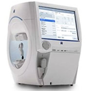
Visual Field:
The visual field test is a subjective measure of central and peripheral vision, or “side vision.” It is used by your doctor to can detect dysfunction in central and peripheral vision which may be caused by various medical conditions such as glaucoma, stroke, pituitary disease, brain tumors or other neurological deficits.
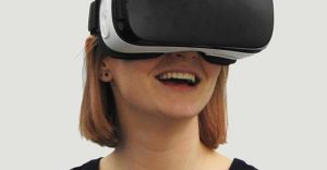
Virtual Visual Field:
We offer virtual visual field testing for patients who are unable to perform the traditional visual field testing due to medical conditions that may prevent them from being able to keep their head steady for a certain period of time. Virtual visual field testing is an alternative that allows for the diagnosing and monitoring of eye diseases
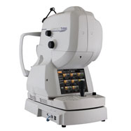
Anterior Segment Imaging:
Anterior segment imaging allows for high resolution photography that allows the doctor to carefully examine the structures of your eyes. The cameras assist in documentation, diagnosis, and patient education.
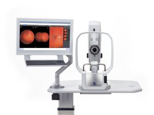
Fundus Photography Imaging:
Fundus Photography is used to inspect anomalies associated with diseases that affect the eye and to monitor the progression of the disease by comparing photographs from one year to the next.
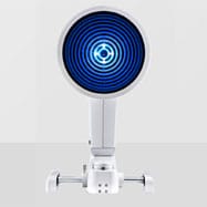
Corneal Topography:
Corneal topography is a computer assisted diagnostic tool that creates a three-dimensional map of the surface curvature of the cornea. The cornea (the front window of the eye) is responsible for about 70 percent of the eye’s focusing power. An eye with normal vision has an evenly rounded cornea, but if the cornea is too flat, too steep, or unevenly curved, less than perfect vision results. The greatest advantage of corneal topography is its ability to detect irregular conditions invisible to most conventional testing.
Corneal topography produces a detailed, visual description of the shape and power of the cornea. This type of analysis provides your doctor with very fine details regarding the condition of the corneal surface. These details are used to diagnose, monitor, and treat various eye conditions. They are also used in fitting contact lenses, fitting orthokeratology lenses and for planning surgery, including laser vision correction.
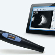
Ophthalmic Ultrasound:
Ultrasonography is a non-invasive instrument used to improve visualization of the structures of the eye in cases in which the Ultrasonography is a non-invasive instrument used to improve visualization of the structures of the eye in cases in which the fundus is obscured from visualization, as in patients with dense cataracts or vitreous hemorrhage. The use of ocular ultrasonography allows the doctor to image the vitreous and retina to detect vitreous degeneration and may also result in earlier detection of ocular melanoma, retinal holes, retinal detachments, and certain optic nerve conditions.
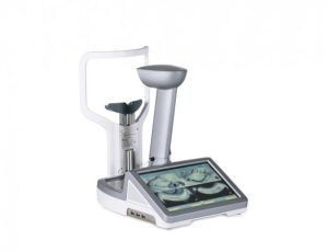
Meibography:
LipiScan meibography is a non-invasive procedure which helps our doctors see and analyze the health of your meibomian glands. It plays an important role in diagnosing and treating the symptoms of dry eye syndrome.
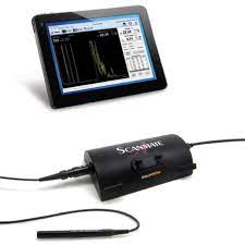
A-scan Biometry:
A-scan biometry, also referred to as A-scan, utilizes an ultrasound device for diagnostic testing. This device can determine the length of the eye which is important in the management of myopia control.

973-248-0060
92 Route 23 N Suite E
Riverdale, NJ 07457
Office Hours
Monday: 9:30am - 6:00pm
Tuesday: 9:30am - 6:00pm
Wednesday: 9:30am - 6:00pm
Thursday: 9:30am - 6:00pm
Friday: 9:00am - 4:00pm
Saturday: 9:00am - 3:00pm
Sunday: Closed
info@riverdalevisioncare.com

